 |
 |
E.A. Glynn, T.P. Quinn, D.P. Fahy, R.E. Dodge, D.S. Gilliam,
and R.E. Spieler
National Coral Reef Institute, Nova Southeastern
University Oceanographic Center (NSUOC),
8000 North Ocean Drive, Dania Beach,
FL 33004
INTRODUCTION:
|
|
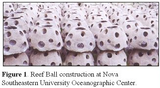
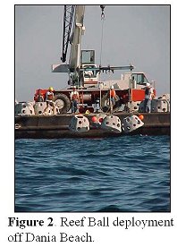
Reef Ball Deployment: In November of 2000, 160 concrete Reef BallTM modules (1.22m wide x 0.9m high) were deployed, at a depth of approximately 15 meters, between the Second and Third Reef tracts off Dania Beach, FL (Figures 1 and 2). The Reef Balls were grouped into 40 quads, with each quad containing four individual Reef Balls. One modified Reef Ball from each quad was designated as the ‘transplant’ ball, and was used as the receptacle for the coral transplants. The other three balls in each quad are part of a more comprehensive study. This multifactorial study is examining the effects of reef structure on fish assemblages, the effects of coral larval attractants on coral recruitment, and the interaction between fish assemblages and coral recruitment. The coral transplants are one such ‘coral larval attractant’ being examined. Coral transplants, and the donor colonies from which they were obtained, are being monitored for growth and survivorship
Study sites:
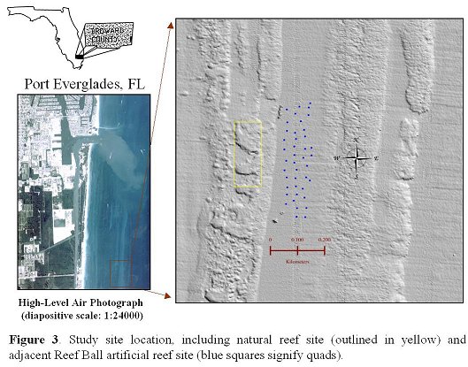 The donor corals and controls of two different sizes are
located at a depth of approximately 10 meters on the Second Reef off Dania Beach
(natural reef site) (Figure 3). The coral transplants are located on the Reef
Balls between the Second and Third Reefs (artificial reef site).
The donor corals and controls of two different sizes are
located at a depth of approximately 10 meters on the Second Reef off Dania Beach
(natural reef site) (Figure 3). The coral transplants are located on the Reef
Balls between the Second and Third Reefs (artificial reef site).
METHODS:
Mapping and Drilling:
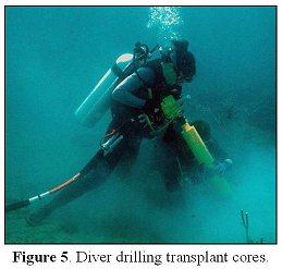
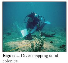
| Forty colonies of two different species of stony coral, Meandrina
meandrites and Montastrea cavernosa, were tagged and mapped at the natural
reef site on the Second Reef (Figure 4). Between March and June 2001, two
core hole plugs with living coral tissue (coral transplants) were obtained
from each donor coral using a hydraulic drill fitted with a four inch core
barrel (Figure 5).
|
|
Transplantation of coral cores and filling
of core holes:
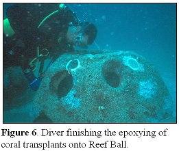 The transplant corals were affixed to the Reef Balls at
the artificial reef site using an underwater adhesive marine epoxy (Figure 6).
One Meandrina meandrites and one Montastrea cavernosa transplant were attached
to each ‘transplant’ Reef Ball.
The transplant corals were affixed to the Reef Balls at
the artificial reef site using an underwater adhesive marine epoxy (Figure 6).
One Meandrina meandrites and one Montastrea cavernosa transplant were attached
to each ‘transplant’ Reef Ball.
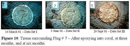
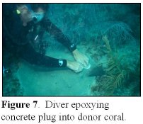 All donor core holes were filled with pre-fabricated,
numbered (1-80), concrete plugs to prevent detrimental effects of bioeroders
(Figure 7). The concrete plug numbers coincide with the transplant numbers
making for ease of comparison of the live tissue surrounding the concrete plug
with the live tissue on the transplant.
All donor core holes were filled with pre-fabricated,
numbered (1-80), concrete plugs to prevent detrimental effects of bioeroders
(Figure 7). The concrete plug numbers coincide with the transplant numbers
making for ease of comparison of the live tissue surrounding the concrete plug
with the live tissue on the transplant.
PRELIMINARY RESULTS:
Monitoring of donor corals:
The 40 donor colonies were photographed using a Nikonos V camera with a 20mm
lens and a 0.75m2 PVC framer marked in 10cm increments, prior to drilling and
after plugging the core holes with the concrete plugs (Figure 8). These colonies
are now being photographed quarterly for the monitoring of health and
survivorship. The donor corals showed 100% survivorship in the second monitoring
session. Additionally, 20 large control corals (comparable size to the donors)
are being monitored at the natural reef site.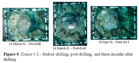
Monitoring of transplants and core hole recovery:
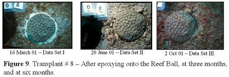
The coral transplants and the core holes are being photographed with a 28mm lens and close-up kit for coral skeleton growth (Figures 9 and 10). Coral skeletal growth is defined as an increase in surface area or linear radius and is being measured quarterly. SigmaScan Pro4 image analysis software (Jandel Scientific Corporation) is being used for all of the photographic analysis. Additionally, 20 small control corals (comparable size to the transplants) are being monitored at the natural reef site.
CONCLUSIONS:
This photographic method is suitable for continuous monitoring and causes no harm to the coral colony. The preliminary results obtained during the first quarterly sample session demonstrated 100% survivorship for all coral plug transplants and donor colonies. It was noted that most coral plug transplants, with exposed skeleton around the margins of the plug, experienced tissue advancement over the bare coral skeleton.
Acknowledgements:
We thank the Broward County Department of Planning and Environmental
Protection, the Florida Marine Research Institute, and the numerous NSUOC
students who helped with the Reef Ball construction and coral transplantation.
| Back to NCRI Projects Page |